America
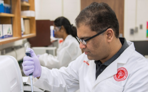
Indian-origin researcher leads study of imaging technology to diagnose lung infections

New York, Aug 16
A study led by an Indian-origin researcher of University of Cincinnati looks to develop a new imaging technique that can identify certain types of lung infections in order to speed up treatment for critically ill patients.
According to Nalinikanth Kotagiri, an associate professor of pharmaceutical sciences at the UC James L. Winkle College of Pharmacy, imaging has the potential to identify specific lung infections in real time -- instead of taking two to three days.
Kotagiri and his team will develop and study the effectiveness of different kinds of injectable probes (metallic contrast agents) that would collect at the site of the infection and immediately light up under a nuclear imaging machine, known as a PET scan.
Currently, radiologists use chest X-rays to confirm the diagnosis of pneumonia and other infections in the lungs, which cannot determine the specifics of the infection or whether the infection is bacterial, viral or fungal.
A specific diagnosis can only be determined by a pathologist, after culturing a sample of lung tissue which is collected from an invasive procedure (called a bronchoscopy) and takes time, typically two-three days.
Critically ill patients, however, such as those with infectious pneumonia and underlying conditions such as chronic obstructive pulmonary disease (COPD), might not have time to spare, says Kotagiri.
"Our solution is to use imaging to identify what is causing the pneumatic episode," within hours, to hasten a treatment plan, he says.
An added benefit, says Kotagiri, is that the contrast agent development process "doesn’t require elaborate processing or preparation timeâ€.
This is critical as development of contrast agents can be time-consuming and complicated. A simple and fast process is expected to reduce preparation time in a clinical laboratory and potentially enable adoption of the technology in a clinical setting.
With this study in animal models, Kotagiri and team will only be looking at bacterial and viral pneumonias in conjunction with COPD, but the imaging approach has the potential to apply to other types of infections such as fungal infections or conditions such as cystic fibrosis.
According to Kotagiri, imaging the patient after treatment could also identify whether the patient is responding to medications such as antibiotics.
Imaging could also potentially aid in determining the right antibiotics to use, targeting the pathogens that were identified, he said.
For the study, Kotagiri has been awarded a five-year $3 million, R01 grant from the National Heart, Lung, and Blood Institute (NHLBI). The R01, or Research Project Grant, provides support for health-related research and development based on the mission of the National Institutes of Health.

26 minutes ago
South Korea, US agree on 'ultimate' goal of denuclearisation of Korean Peninsula

1 hour ago
Together, we will take partnership to new heights: Netanyahu's birthday wishes to 'good friend' PM Modi

1 hour ago
Under your guidance, India achieved impressive results: Putin wishes PM Modi on birthday

1 hour ago
American singer Mary Millben extends heartfelt birthday wishes to PM Modi

2 hours ago
Five-year jail for ex-Custom inspector in Rs 32 crore fraud case in Visakhapatnam

2 hours ago
Unni Mukundan calls playing PM Modi in biopic ‘Maa Vande’ overwhelming and inspiring

2 hours ago
Alia Bhatt wishes PM Modi, hopes his leadership guides India toward greater progress

2 hours ago
Akshay Kumar to PM Modi: Happy Birthday, Captain

2 hours ago
SC cancels bail of Chhota Rajan in hotelier murder case

2 hours ago
'A very happy birthday', Shashi Tharoor greets PM Modi on his 75th birthday

2 hours ago
Kerala HC orders probe into Sabarimala gold plating work after noting discrepancies

2 hours ago
MP to release today female cheetah 'Dheera' in Gandhi Sagar sanctuary

2 hours ago
Chennai Corporation to set up holding facility for aggressive, rabid dogs